


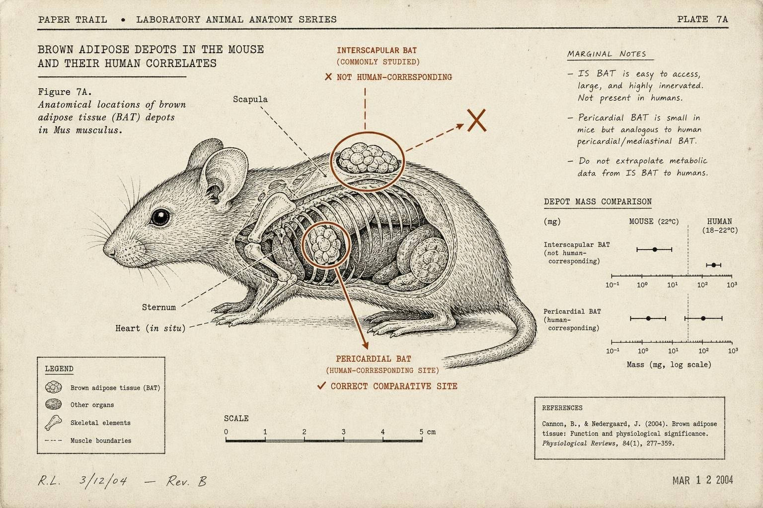

For decades, obesity researchers studying brown adipose tissue — the metabolically active fat that burns energy instead of storing it — have been looking at the wrong body part. A new study in Cell Reports found that the brown fat depot mouse researchers have relied on most, the interscapular pad between the shoulder blades, may not be the one that actually corresponds to the fat humans carry around their collarbones. Instead, the paper traces a heart-associated depot in mice as the better anatomical match for human supraclavicular BAT, the site most studies of human brown fat actually scan (Cell Reports01045-4?rss=yes)). If that holds up, a sizable chunk of mouse-based brown fat research has been mapped onto the wrong tissue entirely — not because mice don't have relevant biology, but because researchers picked the wrong depot to study it in.
This is the quieter version of the mouse-translation problem, and it's more interesting than the usual complaint that "mice aren't people." The failure here isn't species difference in the abstract. It's a specific, correctable anatomical mismatch that nobody checked carefully enough before building a field's worth of assumptions on top of it.
The Mismatch Is Structural, Not Just Biological
The instinct in most science journalism is to treat "mouse models don't translate" as a blanket statement about rodent biology being too different from ours. That's imprecise. The real issue, over and over, is that a specific model — a gene knockout, an anatomical analog, a disease phenotype — gets chosen for convenience and then treated as a stand-in for the human condition without anyone rigorously testing whether the correspondence holds. The BAT depot paper is a case study in exactly that failure mode: researchers used the interscapular pad because it's large, easy to dissect, and has been used since classic 2008 mouse metabolism work, not because anyone confirmed it was the human-relevant site (Cell Reports01045-4?rss=yes)).
Contrast that with a Lab Animal summary of new Duchenne muscular dystrophy mouse models, which took the opposite approach: instead of using a generic dystrophin-deficient mouse and hoping it generalizes, researchers engineered four new strains carrying the exact clinically relevant exon deletions found in actual DMD patients, then confirmed that exon-skipping therapies restored dystrophin production in each one (Lab Animal/Nature). That's a model built to match the human mutation precisely, rather than a model assumed to be close enough. It's also a tell about how much custom engineering it now takes to get a mouse model that regulators and drug developers can trust for a single disease — and that engineering doesn't generalize to the next disease. Every gap gets patched one at a time, at real cost, rather than solved systemically.
When the Patch Is to Stop Using a Mouse's Own Tissue
The most aggressive fix in the current research is to skip rodent biology for the tissue that matters most and transplant actual human cells instead. A Nature report on work from Sergiu Pașca's lab at Stanford describes transplanting human brain tissue into mice that lack a cerebral cortex, producing what the authors call the most extensive integration of human neural tissue into a living animal to date — the grafted tissue grew to fill the vacant space and wired into the mouse's own nervous system (Nature). The appeal is obvious: instead of hoping mouse neurons approximate human neurodevelopmental disease, you put human neurons in a living body and watch what happens. But this swaps one validity problem for another. The tissue is young human brain cells in a fundamentally mouse-shaped circulatory and immune system, and the researchers themselves note the timing of implantation was chosen specifically to avoid the human tissue taking over higher cognitive wiring — a sign of how carefully the chimera has to be constrained to stay interpretable at all (Nature).
The Same Failure Shows Up Outside Biology
A recent Cell review on AI reliability in medicine makes a structurally identical point about a totally different kind of model: predictive systems that perform well in development routinely degrade when deployed into real clinical populations and conditions they weren't validated against, including a widely used sepsis prediction tool that performed worse than chance once predictions made after clinicians had already recognized the condition were excluded (Cell00823-8)). Whether the model is a mouse or an algorithm, the pattern repeats: a system validated under one set of conditions gets treated as generalizable to a different population, and nobody built in the checks to catch the mismatch before it reached deployment.
The fix in both cases isn't more models. It's more scrutiny of whether the model being used actually corresponds to the thing it's supposed to stand in for — before the next decade of papers builds on top of it.